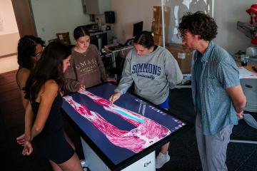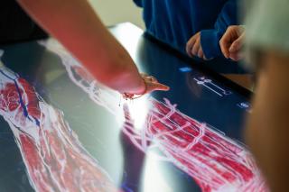
Anatomage Tables are life-size virtual dissection tables that help you learn the structures and systems of the human body. Resembling an operating table, each is a large interactive touchscreen that uses 3D visualization to display fully segmented models from real human cadavers.
Using this technology, you can work collaboratively in small groups to virtually dissect a body. You can examine structures layer by layer without risking damage to specimens. This hands-on approach is particularly helpful if you desire to become a physical therapist, nurse, or allied health professional.
History and Uses of Anatomage Tables
Founded in 2004, Anatomage — a medical equipment manufacturer headquartered in Santa Clara, California — invented the Tables. The company's goal is to “transform science into an accessible, interactive, and experimental journey” for health science majors.
Anatomage Tables are built from full-body scans of real donors, preserving natural anatomical variation. Over 20-plus years, the company has digitally reconstructed more than 2 million anatomical structures.
The system includes a library of more than 1,500 CT and MRI cases with clinical notes, 3D reconstructions, common diseases, traumatic injuries, and rare conditions. You can also review donor medical histories and see how specific diseases affected body systems.
What can you do with an Anatomage Table?
The Anatomage Table helps you understand human anatomy and how structures relate to one another. You can visualize muscles, nerves, blood vessels, ligaments, and organs in three dimensions.
You can create cross-sectional views and animate joints to observe muscle function during movement. This makes the technology especially valuable for physical therapy, sports medicine, orthopedics, and rehabilitation.
As a learning tool, Anatomage Tables offer several advantages as a complement to cadaver dissection, including:
- Repeated practice opportunities
- Better understanding of spatial anatomy and physiology
- Virtual dissection without damaging specimens