
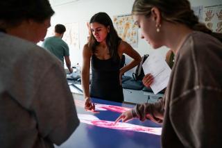
Simmons University has two Anatomage Tables on campus located in the Physical Therapy research lab in Lefavour Hall. This summer, students in the Doctor of Physical Therapy (DPT) program have alternated between lab time with the Tables and the cadaver lab at Harvard Medical School. We spoke to faculty and students about how the new technology enhances their preparation for human dissection.
Two Anatomage Tables are in use on the Simmons campus, allowing students a unique perspective on human anatomy.
“It’s a big computer that looks like an operating table,” says Assistant Professor Steve Wechsler from the Department of Physical Therapy. “The surface is an interactive touch screen, allowing students to touch, move, and maneuver through the bodies represented in the Table.”
The digital bodies represented are based on six donors — real humans who donated their bodies to science — allowing students to navigate different anatomical structures and anomalies therein.
One of the Anatomage Tables was purchased through a generous grant from the George I. Alden Trust. The Tables arrived just before the summer term, allowing new students in the Doctor of Physical Therapy (DPT) program to be among the first to use them, as part of the laboratory component for “Human Anatomy” (DPT 621). When he brings students into the lab, Wechsler helps them navigate the technology.
“The Tables are designed so students can easily toggle on and off different systems,” Wechsler says. “If they are just looking at muscles, they can bring up the musculoskeletal system, or they can focus on gastrointestinal organs in the abdomen. It’s all about what their focus is on a given day.”
Wechsler notes that the Tables complement the work that students do in the Harvard Medical Labs, where they perform cadaver dissection. Simmons’ physical therapy (PT) program is one of the few in New England that offers full cadaver dissection.
“While nothing replaces the experience of performing cadaver dissection, the Tables offer another way for students to interact with incredibly complex material,” says Wechsler.
Complementary Lab Sessions
Students in the “Human Anatomy” course are split into groups of four or five. Each week, they attend three two-hour labs at Harvard and one hour in the Anatomage lab. “In both labs, they are identifying the same structures. The Tables help them to consolidate knowledge and think a bit more clearly and cleanly,” says Wechsler.
As a student with no previous experience with the Anatomage technology, Valeria Caballero ’29DPT is grateful for the opportunity.
“We’ve had time to learn the complexities of the technology,” says Caballero. “It can do such a vast array of things, and Dr. Wechsler has been so supportive. We’ve probably tackled only a small portion of all the functions of that Table! It’s super complex, but Dr. Wechsler has mastered the technology. It’s so interesting to see all the things that it can do.”
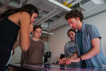
In addition to the scheduled lab times, graduate assistants hold open lab hours, both at Harvard and with the Anatomage Tables on campus. The room is also equipped with whiteboards, encouraging detailed discussion among the groups.
“One group may find a better way to look at something, and the other group can benefit from that discovery. That collaboration is key,” says Wechsler, who acknowledges the learning curve for both the Anatomage technology and human anatomy. “As students learn the technology, they’re better able to navigate the Tables and more skillfully find what they are looking for, and their confidence increases. As they get more comfortable with the technology, I see them make real progress.”
A day in the Anatomage lab can start with a list of items to identify: muscles, nerves, arteries, or any other structure in the body.
“I’m a very visual learner, as are many others in my group,” Caballero says. “You can color code items once you find them, and you can take photos of the Table and refer to those photos while studying.”
The list of assigned structures is easily found on the Tables, allowing students plenty of time for further exploration. “There are only two groups at a time in the Anatomage lab, 10 students maximum, so the instruction is personalized and you can have all of your questions answered. Such a clear vision isn’t always available in the cadaver lab,” she says.
The anatomy lab at Harvard includes about fifty students at a time, though Associate Professor of Practice Rachel Verrill, a coordinator for the Human Anatomy course, is always there to offer guidance to the students. The smaller scale of the Anatomage labs is a benefit for Caballero. “I’ve met all of my professors and feel connected to them, and that’s exactly what I wanted from this program.”
Wechsler notes that some students take naturally to cadaver dissection. Those more sensitive to the experience find that access to the Anatomage technology alleviates anxiety. “The Tables are cleaner, without the same chemical smells of the cadaver lab,” says Wechsler.
Caballero echoes this sentiment, noting, “You don’t need to get scrubs on or smell the formaldehyde. Anatomage is a good break in the week, offering more time to study.”
“Students can work on today’s lab, and review everything they found yesterday at Harvard. The Tables offer another perspective of the same structures, and can even offer a preview of what they will see tomorrow in a real cadaver,” says Wechsler. “It complements what they are doing at Harvard.”
A Unique Look at Human Anatomy
Caballero has found the Tables very useful for identifying muscular structures. “You can transport yourself into a whole array of vasculature, and understand where in the body it is. It’s a base layer of organization that can help you connect the dots when you go into the dissection lab.”
Wechsler notes that the ease of finding structures on the Table allows for further investigation. “Students can map the cardiovascular system and see the spatial relations of what lies next to what structure.”
For fellow student Eytan Pomper ’29DPT, "One of the best aspects of the Anatomage Tables is to see the spatial relations between vessels, muscles, and ligaments. It helps paint a much clearer picture than the Harvard Dissection Lab so that when we do go into the lab, we have a better understanding of their relationships."
Caballero describes another feature of the Tables: you can thinly slice microscopic layers from the top of the body with a swipe of a button, offering new cross-section views.
“In cadaver dissection, students have to work really hard to find a particular structure,” says Wechsler. “The Tables allow time and space to take their critical thinking to the next level. I can ask, what is the function of this system, and if the system is dysfunctional, what do we see?”
For Amanda Goshay ’29DPT, the Tables help students understand the body’s internal systems in a three-dimensional way. “In the Harvard lab, it can be difficult to differentiate muscle layers and make identifications,” says Goshay. “But the Anatomage Tables have features that let you isolate muscles and reposition the body to see it from different angles. This allows us to develop a better understanding of where muscles exist, their functions, and the features they share with other muscles.”
While the donors are not identifiable, the students receive background information on each of the six donors represented in the Anatomage Table, including their age and cause of death. “Each donor provides information on certain pathologies,” notes Wechsler. “One donor died of gastric cancer. You can go into her body and find the cancer, as well as the trauma left by reconstruction surgery.”
There is also a seventh body in the Table that represents a perfect anatomical model, to offer a contrast of what a particular system is supposed to look like. Students can examine the partially collapsed lungs of a donor who died of pneumonia, then switch over to the perfect anatomy example for comparison. Students are allowed to take photographs of these images, for later reference.
“You can also put the bodies in motion,” says Wechsler. “You can look at a hip joint or a knee joint and designate what movement you want it to do, to see what muscles are in motion.”
Expanding Anatomy Learning
While students only have access to the Harvard lab during the summer Anatomy course, the Anatomage Tables will have broader applications throughout their course of study. Wechsler hopes that PT students studying “Movement Science” (DPT 622) in the fall will be able to return to the Anatomage lab for review.
“The clearest use cases are in traditional anatomy and physiology courses, but there are further components of the Tables that could be useful, including real MRI and CT scanned images of real human pathology. This could tie studies of anatomy to pathophysiology and pathoanatomy,” says Wechsler. “Students can explore what they would see during a cardiac event by watching what happens to the heart and what happens on the EKG at the same time.”
Wechsler is confident that they will find further applications for the Tables for PT and nursing students at Simmons.
“It’s very much in line with our programs. Our underlying philosophy is a universal design for learning, which means offering multiple means of engagement to meet students wherever they are, empowering them to access this content,” says Wechsler. “It’s especially vital as we try to meet the needs of more diverse learners, with different experiences and skills.”